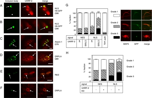
FIGURE 4.
Physiological role of UHRF-2 in pQ pathogenesis. A, fluorescence micrograph of Htt-NLS-Q97 IB surrounded by coexpressed FLAG-tagged UHRF-2 in HeLa cells. IBs are visualized by GFP fluorescence; UHRF-2 was visualized by anti-FLAG and Alexa 546 secondary antibody. Scale bar = 10 μm. Arrows indicate IB; arrowheads indicate neighboring cell nuclei expressing UHRF-2 alone. B, Htt-NLS-Q97 IB transiently expressed in HeLa cells is surrounded by endogenous UHRF-2. Scale bar = 10 μm. Arrows indicate IBs. C, ataxin-1-Q78 IBs transiently formed in HeLa cells colocalize with UHRF-2. IB was labeled with anti-Myc antibody and Alexa 488 secondary antibody. Scale bar = 2 μm. Arrows indicate IBs. D, DRPLAQ80 IBs transiently formed in HeLa cells are positive for UHRF-2. IBs are labeled with anti-FLAG antibody and Alexa 488 secondary antibody. Scale bar = 2 μm. Arrows indicate IBs. E, Htt inclusion is positive for UHRF-2 in vivo. Section from a 6-week-old R6/2 mouse was stained with anti-UHRF-2 antibody. Anti-UHRF-2 antibody stained the IBs. IBs are labeled with anti-ubiquitin antibody. Scale bar = 10 μm. F, DRPLA inclusion is positive for UHRF-2 in vivo. Section from a postmortem DRPLA patient was stained with anti-UHRF-2 antibody. Anti-UHRF-2 antibody stained the IB labeled with anti-ubiquitin antibody. Scale bar = 10 μm. G, UHRF-2 overexpression rescues pQ toxicity in primary cultured mouse neurons. Mouse primary cortical neurons were cotransfected with Htt-Q97 and UHRF-2. After 96 h, coverslips were stained with anti-MAP2 antibody to identify neurons. The shape of the neurons determined by neurite outgrowth was classified into three grades: healthy, fully neurite-grown cells were grouped as grade 1 (a representative cell is shown in the right upper panels); cells with moderately grown neurites were grade 2 (middle panels); cells with unhealthy appearance were grade 3 (lower panels). At least 100 cells from four different coverslips were counted for each transfection. Scale bars = 10 μm. Black bars indicate ±S.E.; *, p = 0.0020; **, p = 0.0407; ***, p = 0.0009 versus wt NLS. H, UHRF-2 knockdown enhances pQ toxicity in primary cultured mouse neurons. Mouse primary cortical neurons were cotransfected with Htt-Q97 and mUHRF-2 or control short hairpin RNA plasmid. After 96 h, coverslips were stained with anti-MAP2 antibody to identify neurons. Evaluation was carried out by the same method as in the overexpression experiment. At least 100 cells from three different coverslips were counted for each transfection. Black bars indicate S.E.; *, p = 0.0049; **, p = 0.0182; ***, p = 0.007 versus NLS RNAi (–).

