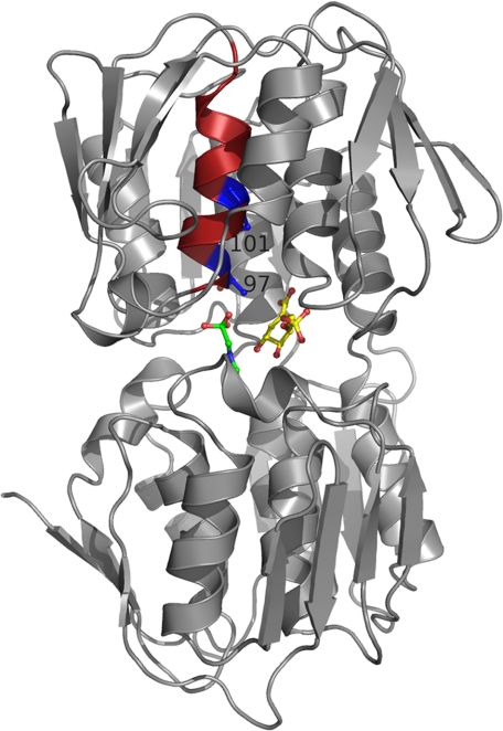
FIGURE 3.
Location of Thr97 and Pro101 in the structure of wild-type EPSPS from E. coli. EPSPS is composed of two globular domains that close upon binding of S3P and glyphosate. The two ligands (shown in yellow and green, respectively) are located in the interdomain cleft of the closed enzyme state. Displayed in maroon is the α-helix in the N-terminal domain containing residues Thr97 and Pro101 (shown in “blue”).

