FIGURE 3.
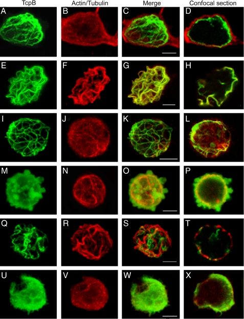
Subcellular localization of TcpB. HEK cells were transfected with pCMV-HA-TcpB plasmid; stained for HA-TcpB (A and E), actin (B), and tubulin (F); and merged (C and G). D and H are sections of C and G, respectively, to indicate the plasma membrane-bound TcpB. To analyze the requirement of microtubules for TcpB localization, cells were treated with nocodazole and stained for TcpB (I) and tubulin (J) and merged (K), and a section of the projected image (L) is shown. To determine the role of the TIR domain for characteristic localization of TcpB, cells were transfected with HA-TcpBΔTIR (M–P) or HA-TcpBTIR (Q–T), stained for TcpB (M and Q) and tubulin (N and R), merged (O and S), and sectioned (P and T). Images U–X are HEK cells expressing HA-TcpBG158A stained for TcpB (U) or tubulin (V), merged (W), and sectioned (X). Scale bars, 5 μm.

