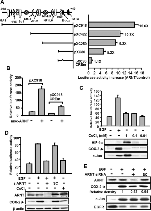
FIGURE 3.
Analysis of ARNT-responsive regions in the 5′-flanking region of the COX-2 gene. A, luciferase vectors bearing various lengths of the COX-2 gene promoter were constructed as indicated. Site-directed mutagenesis of CRE consensus sequences in the 5′-flanking region ranging from –57 to –53 bp of the COX-2 gene was performed. The potential consensus sequences in the 5′-flanking region are indicated. Plasmid transfection was performed as described under “Experimental Procedures.” The luciferase activities and protein concentrations were then determined and normalized. The expression ratios of ARNT (1 μg)-treated cells to control cells are indicated. Results are expressed as means ± S.E. of three independent experiments in triplicate wells for each construct. B, cells were transfected with pXC918, pXC918 CREm, and ARNT expression vector by lipofection. The luciferase activities and protein concentrations were then determined and normalized. Values represent means ± S.E. of three determinations. C, cells were transfected with pXC918 by lipofection. After EGF or CoCl2 treatment for 6 h, the luciferase activities and protein concentrations were determined and normalized. Values represent means ± S.E. of three determinations. Expressions of HIF-1α, COX-2, and c-Jun proteins were analyzed by Western blot analysis using antibodies against HIF-1α, COX-2, and c-Jun, respectively. D, SiHa cells were transfected with pXC918 and ARNT siRNA oligonucleotides by lipofection. After EGF or CoCl2 treatment for 6 h, the luciferase activities and protein concentrations were determined and normalized. Values represent means ± S.E. of three determinations. Expressions of ARNT, COX-2, and β-actin proteins were analyzed by Western blot analysis using antibodies against ARNT, COX-2, and β-actin, respectively. E, OEC-M1 cells were transfected with ARNT siRNA oligonucleotides by lipofection. After EGF treatment for 6 h, the expression of ARNT, COX-2, c-Jun, EGFR, and β-actin was analyzed by Western blot analysis. The relative density of COX-2 protein was quantified as indicated.

