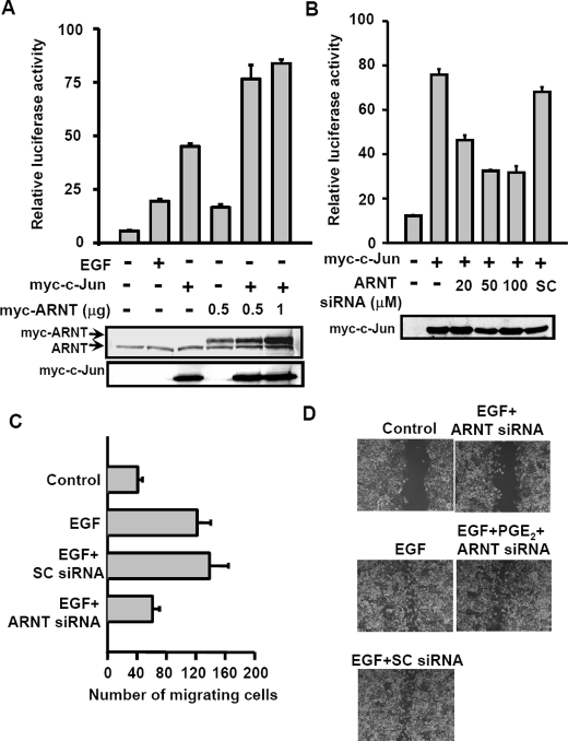
FIGURE 5.
Cooperation between ARNT and c-Jun in promoter activation of the COX-2 gene results in cell migration. A, cells were transfected with pXC-918, Myc-c-Jun, and Myc-ARNT expression vector by lipofection. Cells were treated with or without EGF for 6 h. The luciferase activities and protein concentrations were then determined and normalized. Values represent means ± S.E. of three determinations. Expressions of ARNT and Myc-c-Jun proteins were analyzed by Western blot analysis using anti-ARNT and anti-Myc antibodies, respectively. B, cells were transfected with pXC-918, Myc-c-Jun, and ARNT siRNA by lipofection. The luciferase activities and protein concentrations were then determined and normalized. Values represent means ± S.E. of three determinations. Expressions of Myc-c-Jun proteins were analyzed by Western blot analysis using anti-Myc antibodies. C, cells were transfected with 50 nm ARNT siRNA or scramble oligonucleotides by lipofection. Migration was assessed after 15 h in the presence or absence of EGF. The histogram displays the mean number of migrated cells obtained by counting four separate fields in three independent experiments. Error bars represent means ± S.D. D, cells were transfected with 50 nm ARNT siRNA or scramble oligonucleotides by lipofection. After a wound was made with a pipette tip, cells were treated with 50 ng/ml EGF and 10μm PGE2 for 60 h. The extent of wound closure was observed by using a phase-contrast microscope camera (model DMI 4000 B; Leica).

