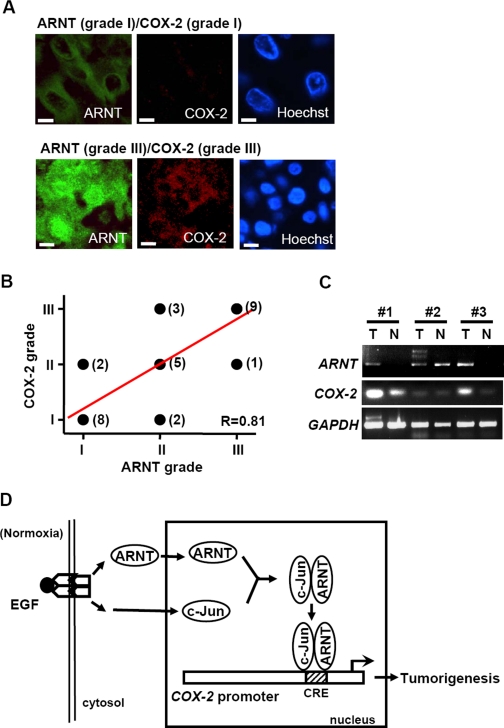
FIGURE 6.
Expression patterns of ARNT and COX-2 in surgical specimens of cervical carcinoma. A, representative pictures of cervical cancer samples at different ARNT/COX-2 grades. The triple-immunofluorescent stain technique was used to identify ARNT (green), COX-2 (red), and nucleus (blue) in cervical cancer tissues. Scale bar, 5 μm. B, a linear correlation (R = 0.81) of ARNT and COX-2 expression in the same surgical specimen of cervical cancer tissues. The case number is indicated in parentheses beside each point. C, total RNA from tumor and normal tissue of different human cervical cancer patients (#1 to #3) was extracted for reverse transcription PCR with COX-2, ARNT, and glyceraldehyde-3-phosphate dehydrogenase primers. D, schematic pathway of COX-2 promoter regulation by EGF stimulation. Activation of EGFR signaling increases nuclear accumulation of ARNT under normoxic conditions. ARNT then interacts with transcriptional factor c-Jun and binds to the CRE site, which results in an increase of COX-2 expression and tumorigenesis.

