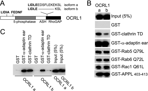
FIGURE 1.
Differential clathrin binding of OCRL1 isoforms. A, schematic view of OCRL1 isoforms a and b. The positions of putative clathrin boxes (LIDIA and LIDLE) and the α-adaptin-binding site (FEDNF) are highlighted in bold. B, GFP-tagged OCRL1 isoforms a and b were expressed in HeLa cells and tested for binding to GST-tagged bait proteins as indicated. Bound proteins were detected by Western blotting with anti-OCRL1 antibodies. C, purified recombinant OCRL1 isoforms a and b were incubated with GST-tagged bait proteins as indicated and bound protein detected by Western blotting with anti-OCRL1 antibodies. TD, terminal domain; α-adaptin ear, α-adaptin appendage domain.

