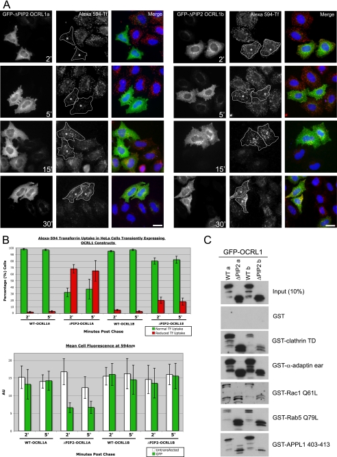
FIGURE 8.
Expression of GFP-OCRL1 isoform a ΔPIP2 impairs transferrin endocytosis. A, GFP-tagged OCRL1 isoform a or b ΔPIP2 deletion mutants were expressed in HeLa cells and Alexa 594-transferrin uptake analyzed at the indicated time points by fluorescence microscopy. Asterisks indicate cells expressing comparable levels of each transfected protein. Bar, 10 μm. B, quantitation of transferrin uptake was monitored in two ways. Top, the level of transferrin uptake in transfected cells relative to neighboring untransfected cells was counted (100 transfected cells counted per construct per time point for each experiment; results are presented as the mean ± S.D. for three experiments). Bottom, the mean transferrin fluorescence per transfected cell was quantitated (10 transfected cells for each construct and time point; results are presented as the mean ± S.D. for three experiments). C, binding of the indicated GFP-tagged OCRL1 constructs to GST-tagged bait proteins was analyzed by Western blotting with antibodies to OCRL1. α-ear, α-adaptin appendage domain.

