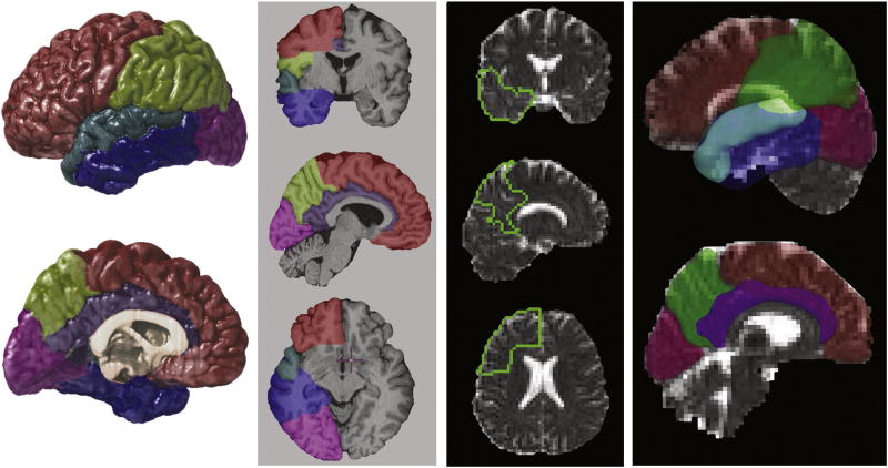
Figure 1.
Regions of interest (ROIs), including the frontal, temporal, parietal and occipital lobe and the cingulate and superior temporal gyrus, were defined in T1-weighted structural MR imaging data (left). A twelve-parameter affine registration method aligned the ROIs to corresponding DTI b0 volumes. The ROIs are shown superimposed on MD images from one subject (right).

