
Figure 7. Amacrine Dm8 neurons receive direct synaptic input from multiple R7 neurons.
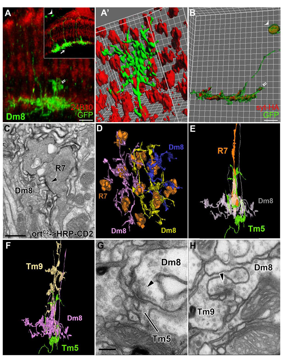
(A–A’) Single Dm8 neuron clones were generated using ortC2-Gal4, hs-Flp, and UAS->CD2>mCD8GFP and visualized with anti-GFP antibody (green). Photoreceptor axons were visualized with MAb24B10 antibody (red). Dm8 neurons extend large processes in medulla stratum M6 (arrow, inset) where they are postsynaptic to 13–16 R7s (A’) and presynaptic to Tm5s. In addition, each Dm8 extends small centrifugal processes to stratum M4 where they are presynaptic to Tm9 (double arrows, A, B). Demonstration of synaptic relations is shown from EM in later panels. (Inset) A low magnification view of (A). The arrowhead and arrow indicate the Dm8 neuron shown in (A). (A’) Isosurface representation of processes of a single Dm8 neuron in a proximo-distal view.
(B) Distribution of presynaptic sites of a single Dm8 neuron. Presynaptic reporter synaptotagmin-HA (red) was localized to the Dm8 processes in strata M6 and M4 (double arrows).
(C) Dm8 is postsynaptic to R7s. A single EM section from stratum M6 shows Dm8 processes marked by an EM marker HRP-CD2 and stained with DAB. R7 terminal was identified based on its vesicle-laden ultrastructure, the presence of capitate projections (not shown) and its location in stratum M6. Presynaptic T-bar ribbon (arrowhead) in R7 profile is juxtaposed to postsynaptic elements of Dm8 with electron-dense membranes.
(D) Serial-EM reconstruction of processes of three Dm8 neurons (pink, yellow and blue) and corresponding R7 terminals (orange). The processes of Dm8 neurons tile stratum M6 with partial overlapping so that each R7 is presynaptic to one or two Dm8 cells.
(E–F) Profiles of R7 (orange), Dm8 (pink), Tm5 (green) and Tm9 (beige) reconstructed from a single medulla column.
(G,H) Single EM sections show that Dm8 is presynaptic to Tm5 (G) and Tm9 (H). Presynaptic T-bar ribbons in Dm8, indicated by arrowheads, point in the presumed direction of transmission.
Scale bar: 5 µm in (A, B); 500 nm in (C); 200 nm in (G) for (G,H)