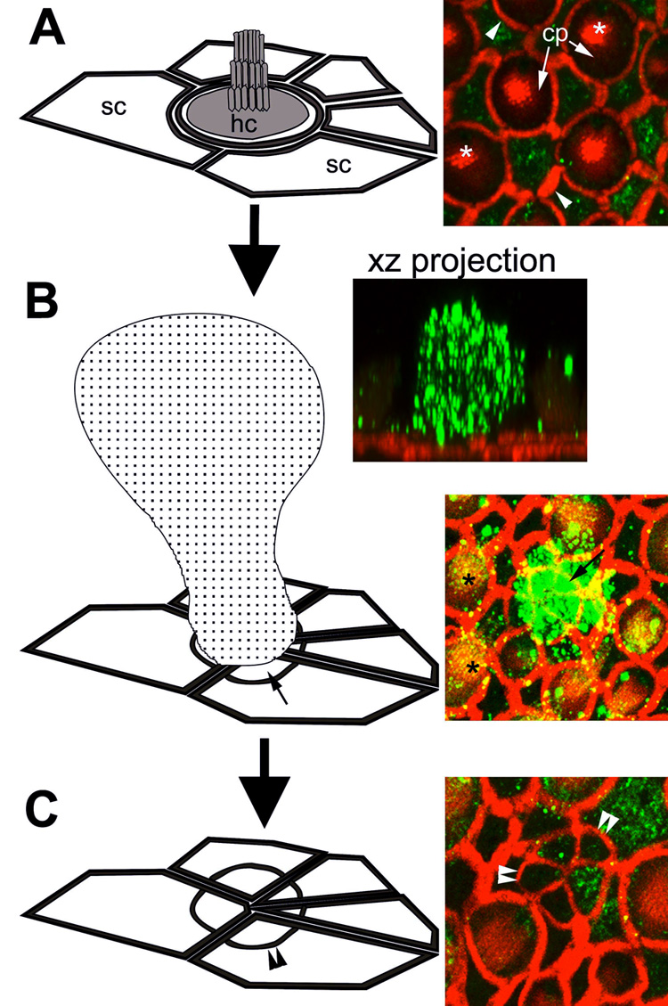
Figure 1.

The morphogenesis of a scar formation. (A) Each saccular hair cell (hc) is surrounded by as many as 6 supporting cells (sc), characterized by their polygonal apical surfaces. In the micrograph on the right, actin (red) labels the actiniferous junctional complexes between adjacent cells (arrowheads), the cuticular plate (cp) and hair bundle (*) (shown in the schematic as shaded regions). Cytokeratins (green) are largely localized in the supporting cells, although some expression in hair cells can be seen surrounding the cuticular plate. (B) The extrusion of a dying hair cell (speckled cell in schematic) correlates with extensive cytokeratin expression within extruded hair cell body (green) in an xz projection. The surrounding supporting cells expand their apical processes (small arrows) to seal the surface of the sensory epithelium, beneath the extruded hair cell. This produces a supporting cell scar formation (C), characterized by a multi-cellular actin ring (double arrowheads). Note the cytokeratin expression (green) within the supporting cell scar. Note also the punctate cytokeratin labeling (yellow-green) associated with hair bundles (*) in (B). A: in situ; B and C 24 hours after explantation. (Not to scale, for illustrative purposes only.)
