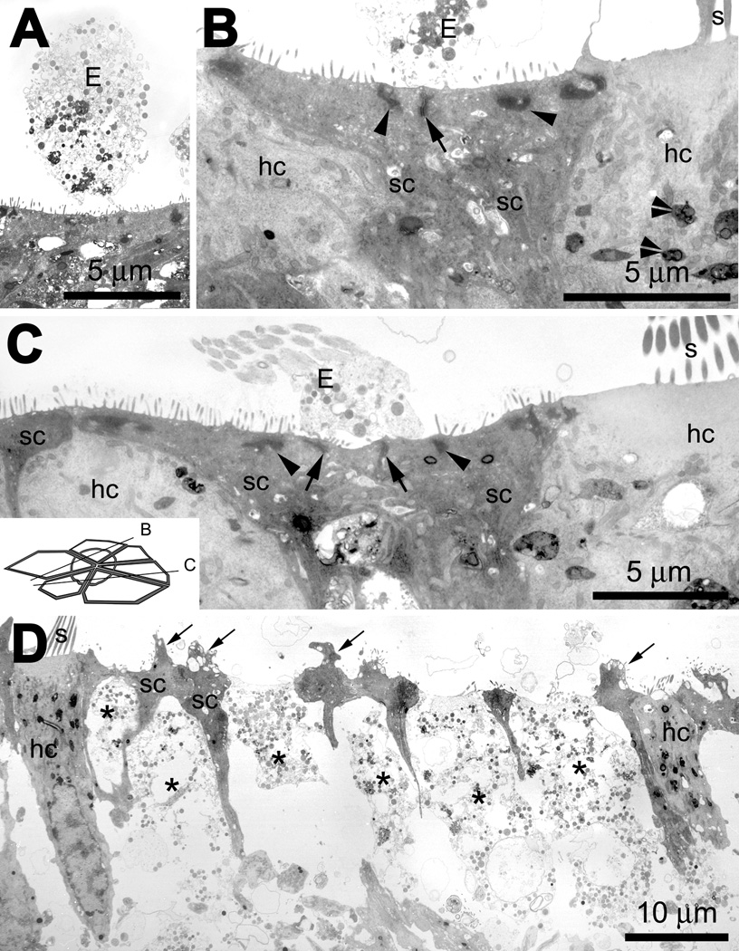
Figure 11.

(A) A low resolution image of an extrusion (E) with cytoplasmic contents above the sensory epithelium of an explant treated with gentamicin only. (B) In a gentamicin only-treated explant, extruded cellular material (E) is located above a sectioned scar formation; the expanded apices of two supporting cells (sc) meet and form a junctional complex (including tight junctions; arrow), with islands of electron-dense microfilamentous material (arrowheads) near the luminal membrane. Surviving hair cells (hc) contain electron-dense multi-vesiculated “gentamicin” bodies (double arrowheads). (C) Another example of a cross-sectioned supporting cell scar formation, where the expanded apices of three supporting cells (sc) forming two junctional complexes (including tight junctions, arrows) beneath extruded cellular material (E). There are also islands of electron-dense, microfilamentous material (arrowheads) near the luminal membrane of these supporting cells. Inset shows approximate cross-sectional planes for panels B and C. (D). In gentamicin and cytochalasin D-treated explants, hair cells (hc) that survived drug administration are similar in density to hair cells in gentamicin-treated explants, with well-defined stereocilia protruding from the cuticular plate. Remnants of dead hair cells are emplaced within the sensory epithelium and highly electron-translucent (*), with little cytoplasmic material beneath the fragmenting cell apex. The supporting cell apices retained their junctional complexes, and often had cytoplasmic and vacuolated protrusions into the extracellular space (arrows).
