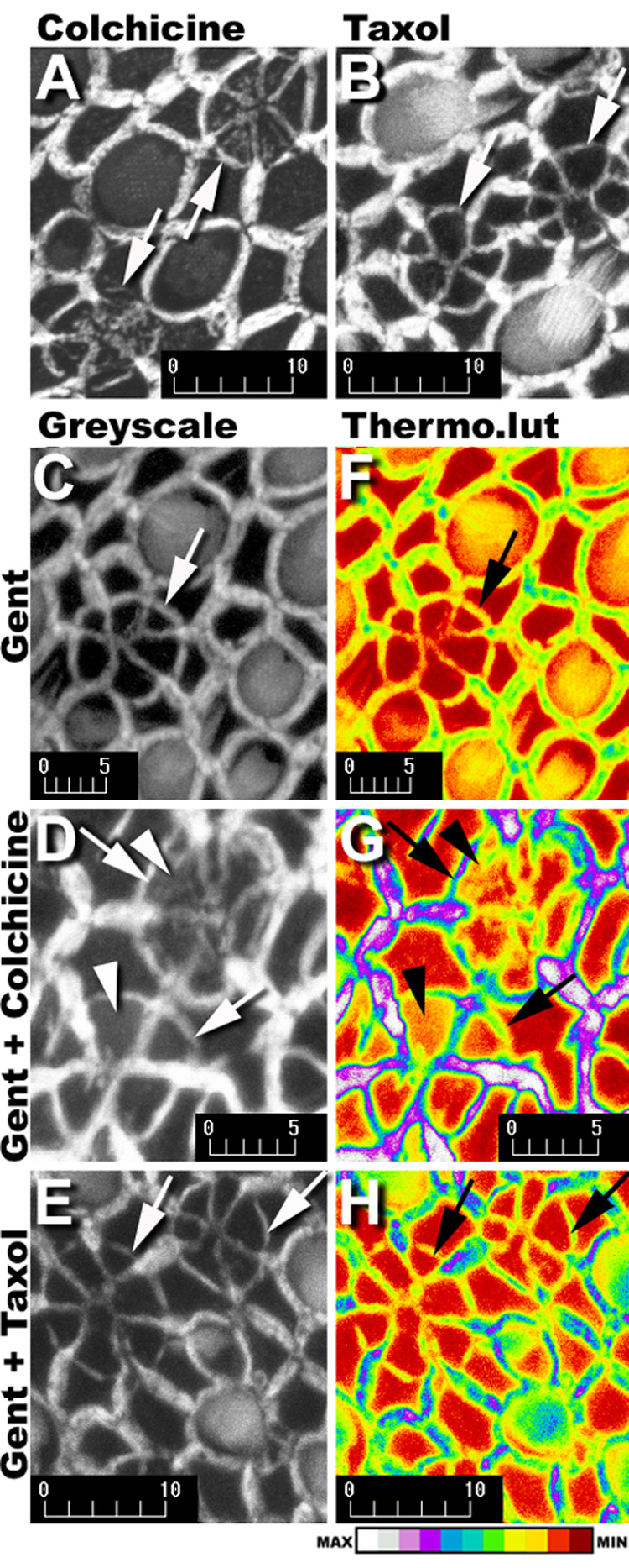
Figure 4.

Phalloidin-labeled multicellular rings/cartwheels (↑) in explants treated with gentamicin plus colchicine (A) or gentamicin plus taxol (B). In colchicine-treated explants (A, D), deposition of F-actin (arrowhead) occurs within the expanded apical processes of supporting cells participating in the scar formation, but not in scar formations of gentamicin treated (C), or gentamicin plus taxol-treated explants (B,E). When the grayscale images in C–E are loaded with the Thermo look-up table (F–H), a dark red indicates the absence of phalloidin-conjugated Alexa 568 fluorescence, and white represents maximum fluorescence intensity (see intensity bar at bottom). Supporting cell apices not participating in scar formations in all explants (F–H), are red or dark red. Colchicine-treated explants (G) have yellow pixels within the central region of the expanded supporting cell apices participating in scar formations (arrowheads) in contrast to the dark red pixels throughout the scar formations in explants treated with gentamicin only (F) or gentamicin plus taxol (H). Scale bars in microns.
