
Figure 8.
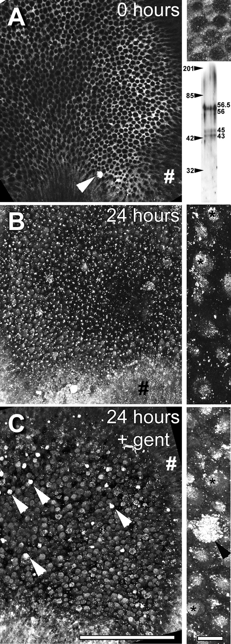
(A) Bullfrog saccule (from right ear) fixed immediately after otolithic membrane removal and immunolabeled for cytokeratins. At low resolution, labeling is distinctly localized in supporting cell apices, outlining the round apices of hair cells. Note the intensely labeled extrusion near the growth zone (arrowhead). Upper inset on the right shows higher resolution projection image (of several focal planes) from the same saccule, with the labeling preferentially localized in the polygonal apices of supporting cells, and also as puncta in the round hair cell apex. Lower inset shows a Western blot of three distinct protein bands at 43, 45 and 56/56.5 kD for the cytokeratin antibody. Figures on the left indicate the position of the molecular weight markers. (B) Left saccule incubated in ACM only for 24 hours reveals loss of the distinctive cytokeratin immunolabeling from supporting cell apices, and the appearance of bright puncta throughout the sensory epithelium, with generalized labeling of cells in the sensory epithelium. Inset on the right: Higher resolution projection image reveals preferential cytokeratin immunolabeling of round hair cell apices over adjacent supporting cells, with intense punctate labeling associated with the hair bundle (*), and elsewhere. (C) Right saccular explant treated with gentamicin for 6 hours and allowed to recover in ACM for 18 hours, reveals significant cytokeratin immunolabeling in the round hair cell apices. Note several intensely-labeled cells, probably extrusions (arrowheads). Inset on the right: Higher resolution projection reveals intense labeling in an extrusion and in many hair cell apices and bundles (*). Note the labeling of the transitional epithelium (#) in all low resolution images. Scale bars = 200 µm in main images and 10 µm in insets.