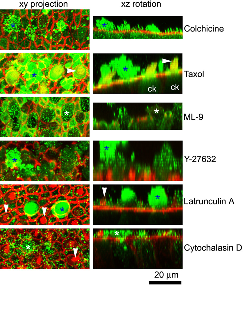
Figure 9.

Cytokeratin-labeled hair cell extrusions (*) are present in all explants, though far less frequently in latrunculin A and cytochalasin D-treated explants after gentamicin treatment. Phalloidin labeling for F-actin (red) indicates the borders of supporting cells and hair cells, some with their stereociliary bundles (arrowheads). The left column shows a projection of the optical planes incorporating the extrusion. The right column shows a 3-D rotation of the stack shown in the left column, indicating the extrusion (*). Note the cytokeratin labeling in hair cells (ck) in the taxol xz rotation panel. Numerous fluorescent speckles (green) also occurred above the sensory epithelium.
