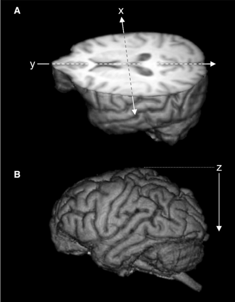
Figure 1. Three-Dimensional Reconstructions of Magnetic Resonance Images of a Representative Chimpanzee Brain.

(A) Illustrated is a three-dimensional-rendered MR image of chimpanzee brain cut in to reveal the axial view. “x” and “y” indicate the orthogonal planes (sagittal and coronal, respectively) referred to in Figure 2. Arrow directions refer to ascending slices displayed in Figure 2.
(B) The z axis indicates the axial plane referred to in Figure 2.
