
Figure 1. Cytotoxic effect of the MEK inhibitor CI-1040 in a panel of melanoma cell lines.
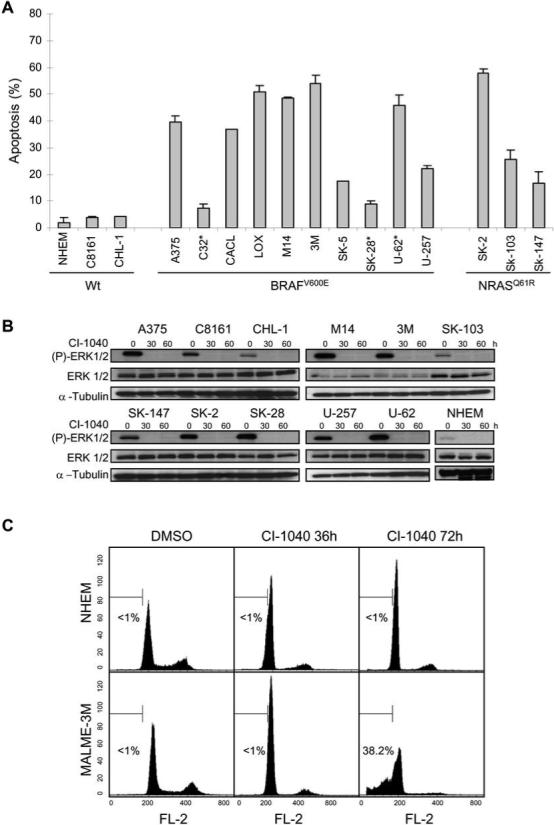
(A) Apoptotic sensitivity of normal human epidermal melanocytes (NHEM) and melanoma cell lines to the MEK inhibitor CI-1040. Cells were treated in triplicate with either DMSO as a control or 2 μM CI-1040 for 72 h and apoptosis (% sub G1 fraction) was determined by cell cycle analysis using flow cytometry. Data was normalized to the control and expressed as the mean ± SEM. Cell lines are grouped according to BRAF or NRAS mutation status. Astericks (*) indicate PTEN mutation. (B) Western blot analysis of phosphorylated ERK1/2 (P-ERK 1/2), and total ERK 1/2 expression in NHEM and melanoma cell lines treated as in (A). Whole cell lysates (30 μg each) were subjected to Western blotting using the indicated phospho-specific antibodies to detect activation of the kinases. The blots were re-probed with their respective total protein antibodies. The blots were re-probed with α-tubulin as a loading control. (C) Induction of G1 arrest in NHEM (top panels) and apoptosis in MALME-3M melanoma cells (lower panels) by CI-1040. Response of cells to DMSO (left panels) or CI-1040 for 36 hours (middle panels) or 72 hours (right panels) was analyzed by flow cytometry. A percentage of apoptosis quantified independently is indicated in each histogram.