Figure 4.
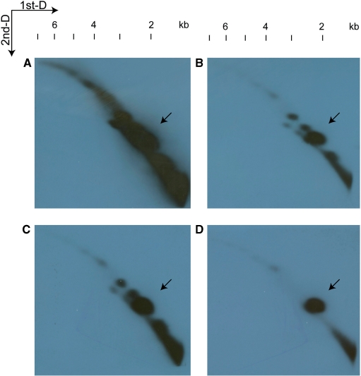
Patterns of minicircle DNA resolved in 2D-gel. Whole-DNA extract from H. triquetra was subjected to 2D-gel as described in the Methods section. The first dimension is represented horizontally (left to right) and the second dimension vertically (top to bottom). Minicircle DNA were detected with 1.1-kb (A), 0.4-kb (B) and 0.6-kb (C) psbA NCR, and 0.7-kb psbA gene fragment (D). Four images were aligned with respect to their DNA migration in both dimensions of electrophoreses. The putative psbA minicircle spots indicated by the arrows were perfectly aligned. The sizes of hybridization signals can be revealed by the size ladder on the top.

