Figure 5.
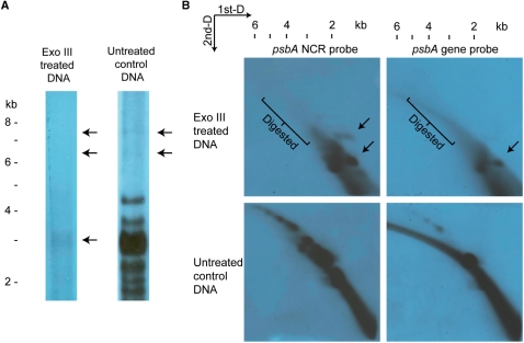
Exo III-treated minicircular DNA revealed after PFGE and 2D-gel electrophoresis. Exo III-treated whole-DNA extract from H. triquetra was subjected to PFGE (A) and 2D-gel (B), and detected with 1.1-kb psbA NCR fragment [and psbA gene fragment in (B) only]. (A) The 6–8 kb minicircular DNA bands (APBs) were removed in the Exo III lane, while the putative 3-kb psbA minicircle band was diminished in intensity. (B) Hybridization signals of larger than 2 kb were removed after Exo III treatment as revealed in both 2D-gels detected by psbA NCR and gene fragments. Extra spots indicated by the arrows were observed in these two images when comparing with the controls. DNA markers were linear DNAs from a commercial source (Invitrogen Corporation).

