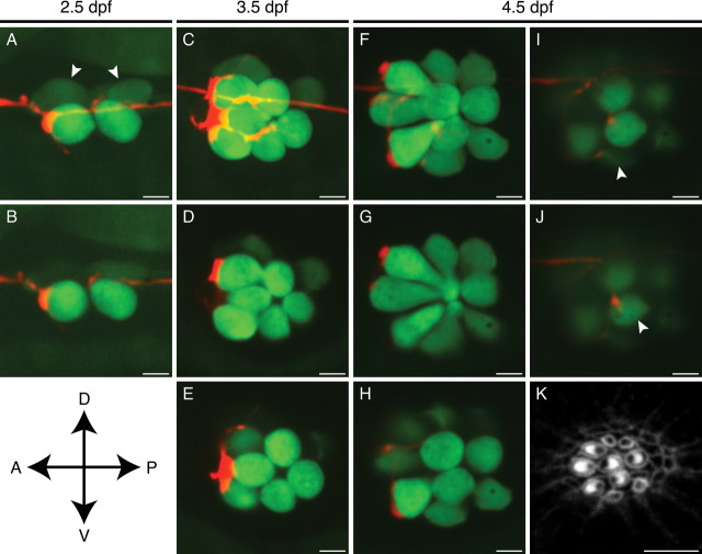
Figure 2.
In vivo imaging of afferent synaptogenesis in a developing neuromast. A, In a maximal-intensity projection of a Z-stack of an anteroposterior neuromast at 2.5 dpf, an mCherry-labeled afferent fiber (red) forms a putative synapse with the rostralmost of the hair cells expressing GFP (green). Two immature hair cells are only dimly labeled with GFP (arrowheads). B, A selected confocal section of the neuromast in A shows the extensive contact between the terminal and one hair cell as well as a substantially smaller contact with a second. C, A maximal-intensity projection of the same neuromast at 3.5 dpf illustrates extensive neuronal contact with the three posteriorly polarized hair cells. D, E, Selected optical sections of the neuromast depicted in C delineate the individual contacts. F, A maximal-intensity projection of the same neuromast at 4.5 dpf demonstrates five putative synapses, of which four occur with posteriorly polarized hair cells. G, H, Large boutons have formed on the three largest posteriorly polarized hair cells. I, A newly formed hair cell has been innervated (arrowhead) just as its hair bundle has begun to polarize posteriorly (see K). J, One innervated hair cell of this neuromast (arrowhead) is of the opposite polarity with respect to the others (see K). K, Staining of hair bundles in this neuromast with fluorescent phalloidin reveals the polarities of the hair cells at 4.5 dpf. The stereocilia in each bundle display a crescentic pattern of fluorescence surrounding a dark spot at the site of the kinocilium. A, Anterior; P, posterior; D, dorsal; V, ventral. Scale bars, 5 μm.

