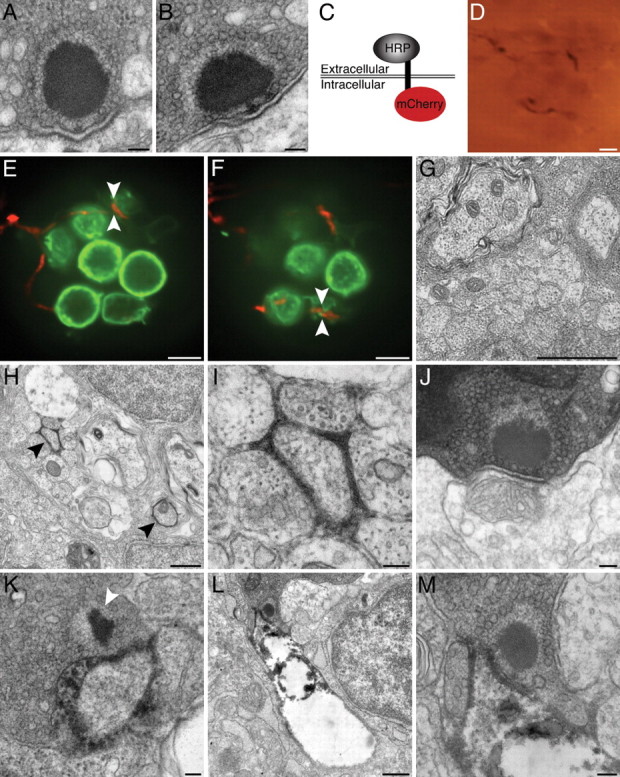
Figure 5.

Correlative electron microscopy with HRP-mCherry. A, A ribbon synapse in a 2 dpf wild-type larva is indistinguishable from those in older animals. B, The synapse in a 5 dpf wild-type larva exhibits the characteristic features of a ribbon synapse, including a presynaptic dense body or ribbon, a halo of tethered synaptic vesicles, and prominent presynaptic and postsynaptic densities. C, Expression of the HRP-mCherry protein in the neurolemma places the fluorescent mCherry component intracellularly and the HRP moiety extracellularly. D, A bright-field micrograph depicts an afferent terminal expressing HRP-mCherry within a neuromast. The densely labeled fiber, which is also depicted in E, F, and H–M, is visible through the plastic resin in which the specimen has been embedded. E, An optical section through a neuromast of a living Brn3c:gap43-GFP larva features hair cells expressing a membrane-localized form of GFP (green). An afferent fiber labeled with HRP-mCherry (red) innervates three of the hair cells. The region bracketed by arrowheads is examined in greater detail in K. F, In an optical section through the basal region of the same neuromast, arrowheads bracket a site that was later explored under the electron microscope (L, M). G, A transverse section of the PLL nerve in a wild-type 5 dpf larva demonstrates several afferent axons. H, In a transverse section through a PLL nerve, two afferent fibers that express HRP-mCherry (arrowheads) produce prominent electron density in the surrounding extracellular space. The weakly labeled fiber in the lower right did not innervate the neuromast depicted in D–F and H–M. I, A higher-magnification view of the labeled neuron at the top left of H illustrates a localized precipitate that does not damage nearby cells. J, An unlabeled afferent neuron lacking electron density synapses with a hair cell of the neuromast. K, A synaptic ribbon (arrowhead) in the region of membrane contact denoted by arrowheads in E verifies that the membrane contact observed by light microscopy represents an afferent synapse. L, This ribbon synapse occurs at the site of membrane apposition bracketed by arrowheads in F. In this instance, the neuron has become distorted and exhibits poor preservation of intracellular organelles. M, Viewed at higher magnification, the ribbon synapse in L illustrates the typical attributes of hair-cell afferent synapses. Scale bars: A, B, I–K, M, 100 nm; D–F, 5 μm; G, H, L, 500 nm.
