
Figure 6.
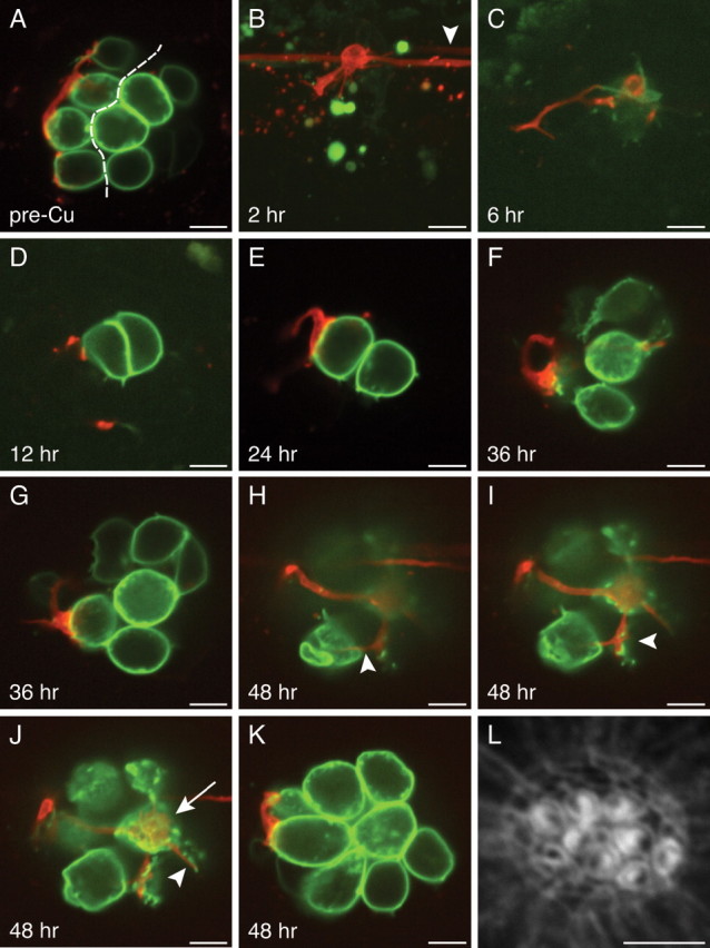
Reinnervation of regenerated hair cells. A, In an optical section through a 3 dpf neuromast before hair-cell elimination, the axis of planar cellular polarity (dashed line) can be inferred from the positions of the constituent hair cells. The afferent fiber has selectively synapsed with posteriorly polarized hair cells. B, In a maximal-intensity projection of the same neuromast 2 h after the application of 10 μm Cu2+, the hair cells have been eliminated, and the neuron has retracted its terminals. Note the presence in the lateral-line nerve of another labeled neuron that does not innervate this neuromast (arrowhead). C, After 6 h, the neuromast contains one weakly fluorescent progenitor that has not yet undergone mitosis to form two new hair cells. D, Twelve hours after Cu2+ treatment, the newly formed posteriorly polarized hair cell receives a small synapse. E, By 24 h, the synapse depicted in D has grown in size and in the extent of membrane contact. F, G, At 36 h after treatment, the neuron appears to contact two or three hair cells, but their polarities cannot be inferred because of the complex organization of the neuromast. It is likely, however, that the synapse depicted in G is identical to that in D and E. H–K, By 48 h, the neuromast has grown to encompass eight mature hair cells with polarized hair bundles (see L). These four panels are ordered from the bases to the apices of the hair cells. H, A thin neurite reaches an anteriorly polarized hair cell (arrowhead). I, A larger bouton contacts the ventralmost of the posteriorly polarized hair cells (arrowhead). J, A synaptic contact blankets the basal surface of a posteriorly polarized hair cell (arrow), whereas only a tenuous process reaches an anteriorly polarized hair cell (arrowhead). K, The afferent neuron forms voluminous boutons on two posteriorly polarized hair cells. L, Staining with fluorescent phalloidin 48 h after treatment defines the polarities of the 10 hair bundles. Scale bars, 5 μm.