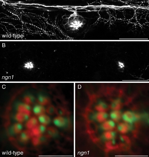
Figure 7.
Hair-cell polarity in the absence of innervation. A, A maximal-intensity projection of a confocal Z-stack depicts immunolabeling for acetylated α-tubulin in the lateral line of a 5 dpf wild-type larva. The PLL nerve and superficial sensory neurons are labeled, as well as microtubules in the apices of hair cells. B, Immunolabeling of a neurogenin1 mutant sibling for acetylated α-tubulin illustrates the absence of a PLL nerve. Labeling persists in the microtubules of hair cells. C, Staining of a wild-type neuromast with fluorescent phalloidin (red) and immunofluorescent labeling of acetylated α-tubulin (green) reveal the polarities of the hair bundles in this anteroposterior neuromast. D, The hair-bundle polarities of a neurogenin1 mutant neuromast are unperturbed despite the lack of innervation. Scale bars: A, B, 30 μm; C, D, 5 μm.

