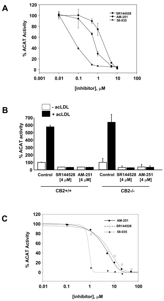
Fig. 3.
AM-251 and SR144528 inhibit the stimulation of cholesterol esterification by acLDL independent of CB2 expression. (A) Raw 264.7 macrophages were pretreated with increasing concentrations of AM-251, SR144528 or 58-035 for 1h prior to supplementation with acLDL (100 μg/ml) and [3H]oleate (1 μCi/ml) as indicated. Following a 6 h incubation, the amount of cholesteryl [3H]oleate formed was determined and the ACAT activity expressed as percentage of the mean cholesteryl [3H]oleate dpm/mg realized for controls (vehicle only). Each experiment was performed independently twice, each with triplicate samples. (B) AM-251 and SR144528 inhibit acLDL-stimulated cholesterol esterification in resident peritoneal macrophages from wild type and CB2 null mice. Wild type (CB2+/+) and CB2 null (CB2−/−) resident macrophages were pretreated with AM-251 or SR144528 for 1h prior to the addition of acLDL (100 μg/ml) and [3H]oleate (1 μCi/ml). After 6 h, total lipids were extracted and the amount of cholesteryl [3H]oleate formed was determined. ACAT activity is expressed as the percentage of the mean cholesteryl [3H]oleate dpm/mg realized for controls (vehicle only). Each experiment was performed independently twice, each with triplicate samples. (C) Mouse liver microsomes were preincubated for 30 min with various concentrations (0–20 μM) of the inhibitors as indicated. [14C]Oleoyl-CoA (0.5 μCi) was added and the amount of cholesteryl [14C]oleate formed after a 5 min incubation at 37°C was determined. ACAT activity is expressed as the percentage of cholesteryl [14C]oleate formed in the absence of inhibitors ± SD for triplicate samples. In some cases the error bars are smaller than the symbol and are not visible.

