
Fig. 1.
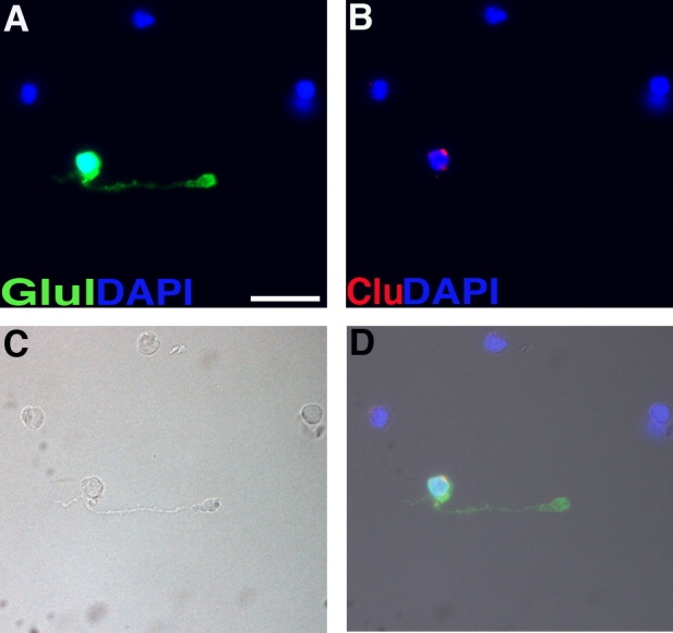
Prospective identification of single Müller glial cells by morphology. Micrograph of cells dissociated from an adult mouse retina. A: Immunostaining for glutamine synthetase (Glul). B: Dissociated cell in situ hybridization for clusterin. C: Brightfield image. D: Merged image. Scale bar = 10 μm in A (applies to A–D).