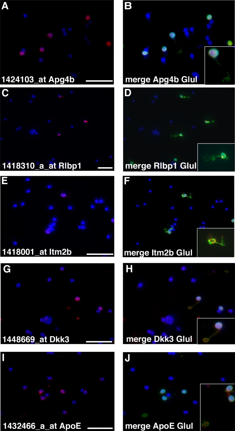
Fig. 6.

Dissociated cell ISH confirms expression in Müller glia. A–J: Retinae from mature mice were dissociated, plated on slides, and processed for detection by ISH for the indicated probes (ApoE, Dkk3, Apg4b, Itm2b, Rlbp1). Merged images show DAPI stain and immuno-staining for Glul and ISH. Insets are digital magnifications. Scale bar = 25 μm in A (applies to A,B), C (applies to C,D), E (applies to E,F), G (applies to G,H), and I (applies to I,J).
