Figure 5.
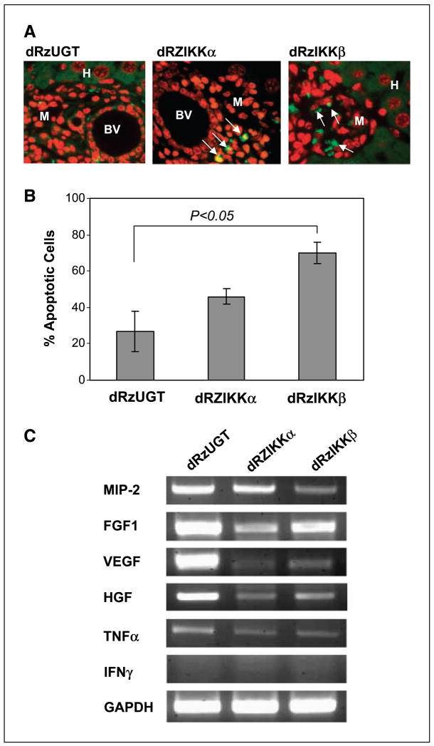
dRzIKK induction of cellular apoptosis. A, paraffin-embedded liver tissues with micrometastatic Ras-melanoma were subjected to DeadEnd Fluorometric TUNEL assay for detection of apoptosis. The TUNEL-positive cells are visualized in green fluorescence (arrow) in a red propidium iodide background by fluorescence microscopy. Data are from one of two independent experiments showing similar results. BV, blood vessel; M, melanoma cells; H, hepatocytes. B, Ras-melanoma cells (5 × 10⁶) were transfected with either dRzIKKα, dRzIKKβ, or dRzUGT. Thirty-six hours after transfection, the apoptotic cells were examined using TUNEL assay and analyzed by flow cytometry by measuring fluorescein-12-dUTP at 520 nm. C, dRzIKK regulated expression of angiogenesis factors. Ras-melanoma cells (5 × 10⁵) were i.v. injected into C57/BL6 mice (three mice/group) for 3 days, and subsequently, 100 μg vector DNA of dRzIKKα, dRzIKKβ, or dRzUGT was delivered i.v. Three days after dRzIKK injection, mice were euthanized and the livers were homogenized. The total RNA was extracted and subjected to RT-PCR assay for MIP-2, FGF1, VEGF, HGF, TNFα, and IFNγ. GAPDH was used as a loading control.

