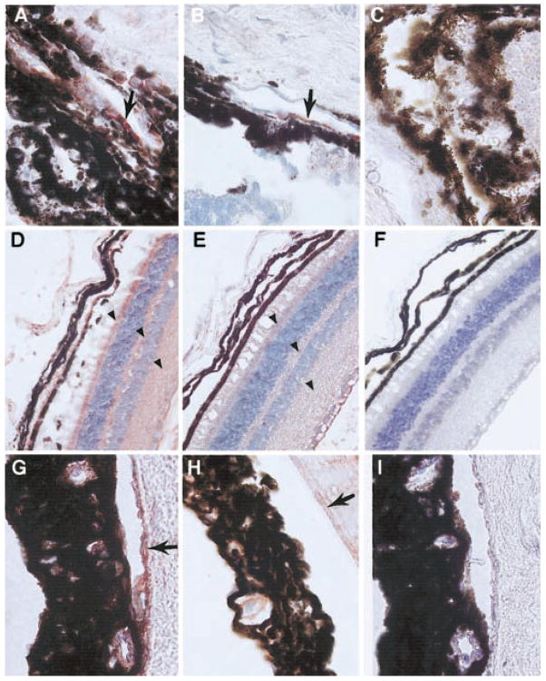
Fig. 4.
Immunolocalization of mDuffy and CXCR2 in the eye of transgenic mice treated with MIP-2. Immunoreactive CXCR2 (red staining) was observed in the endothelial cells of capillaries and blood vessels near the limbus, apparently involved in the angiogenic response to MIP-2 in transgenic mice (A) and WT mice (B), compared with control sections stained with a nonspecific IgG (C; original magnification, 125×). In transgenic mice, mDuffy immunoreactivity was stronger in the endothelial cells of the choroid, in the cells of the neural retina, particularly the rods and cones, in the cell processes comprising the outer plexiform layer, and the inner plexiform layer (D), compared with WT mice (E) and control treated with nonspecific IgG (F; original magnification, 50×). The blood vessels of the iris stain positively for immunoreactive mDuffy in transgenic mice (G) and to a lesser extent, in WT mice (H), and control tissues treated with a nonspecific isotype-matched IgG exhibit no immunoreactivity (I; original magnification, 125×). Arrows indicate positive staining for CXCR2 (A and B) and Duffy (D, E, G, and H).

