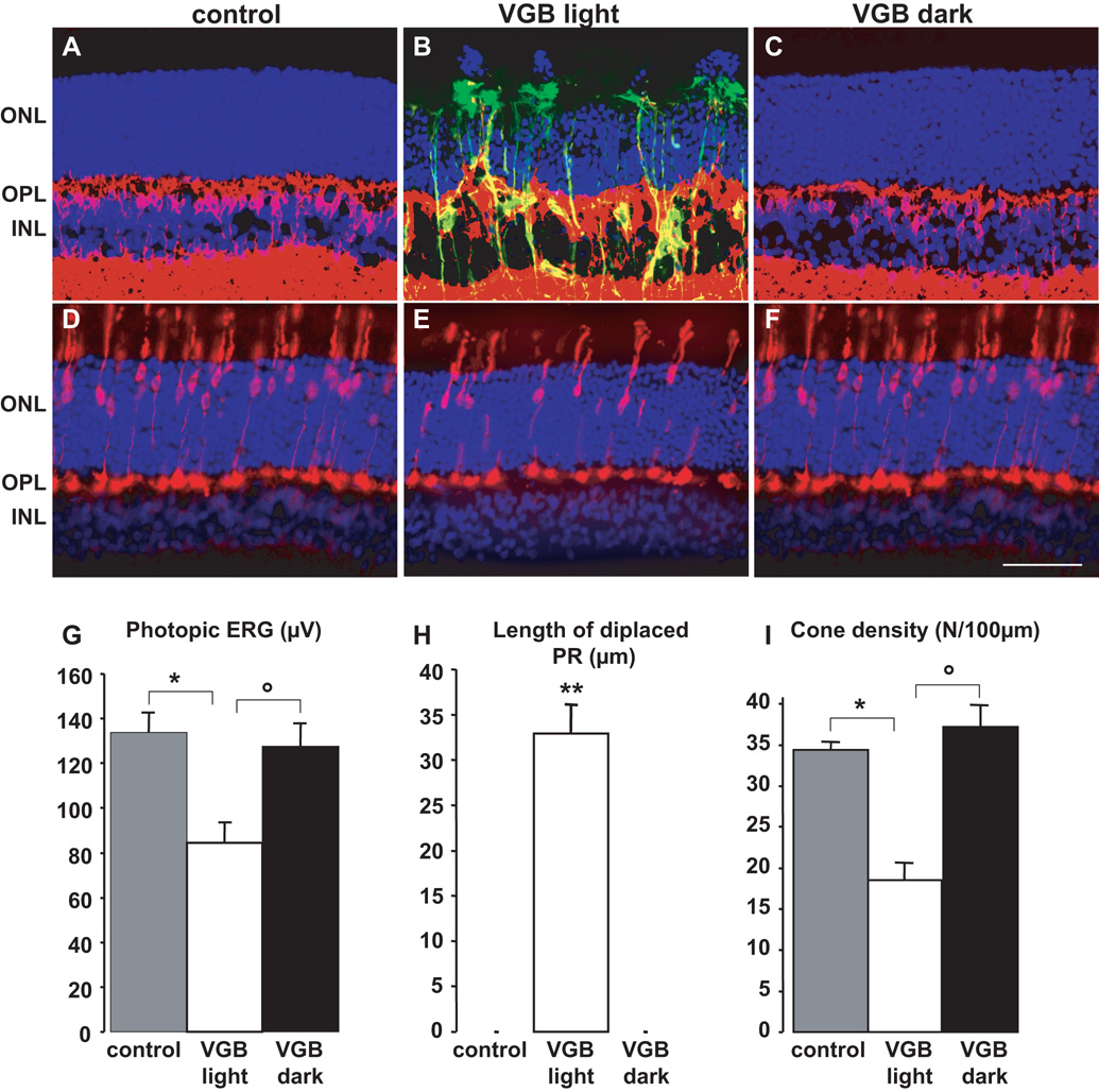
Figure 1. The in vivo retinal phototoxicity of vigabatrin.
(A–F) Retinal sections showing the lesions in a rat treated with VGB for 45 days in room light (B, E, VGB light) and absence of lesions in a VGB-treated rat maintained in darkness (C, F, VGB dark) and in a control animal (A, D). Sections were stained with the nuclear dye, DAPI, (blue in A–F) and immunolabelled for GFAP (green in A–C), Goα (red in A–C) and cone arrestin (red in D–F). Photoreceptor nuclei displaced above the outer nuclear layer (ONL), Goα-positive bipolar cell dendrites sprouting into the ONL and GFAP-positive processes extending vertically throughout the retina are only observed in rats treated with VGB in the 12h/12h light/dark cycle (B), not in control animals (A) or VGB-treated animals maintained in darkness (C). Similarly, fewer cone arrestin-positive photoreceptors and their inner/outer segments were observed in areas with normal retinal layering in VGB-treated rats exposed to a 12h/12h light/dark cycle (E), than were observed for control (D) or VGB-treated rats maintained in the dark (F). Quantification of photopic ERG amplitudes (G), lengths of retinal areas with displaced photoreceptor (PR) nuclei (H) and cone inner/outer segment density (I) in control rats (s.e.m., n=9), for VGB-treated animals either exposed to a 12h/12h light/dark cycle (VGB light, s.e.m., n= 10) or maintained in darkness (VGB dark, s.e.m., n= 10). The scale bar represents 50µm (IPL: inner plexiform layer).

