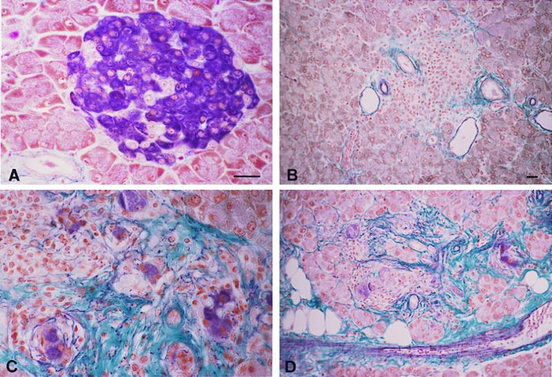
Figure 2.

A severe pancreatic fibrosis develops in TNF/CD80 mice with a mutated TNF-LTα locus. Seven micrometer-thick pancreatic paraffin sections were stained with the aldehyde-fuchsin method to reveal insulin cells (in violet) as well as collagen and elastin fibers (green and violet, respectively). (A) Normal mouse islet; B–D, mouse islets from three different transgenic animals bearing the TNF-LTα mutation. B shows an atrophic islet in which no β cells remain; only infiltrating cells are seen, and the fibrotic reaction remains moderate, as usually observed in TNF/CD80 nonmutant transgenic mice. In C, several clusters of isolated β cells (violet) are surrounded by fibrosis (green), together with lymphocytes (Upper Left); unaffected adjacent exocrine cells are also seen (Upper Right). In D, fibrosis extends to the adjacent exocrine tissue, which is progressively replaced by adipocytes (white holes). Bars = 20 μm (same magnification in A–C and in B–D).
