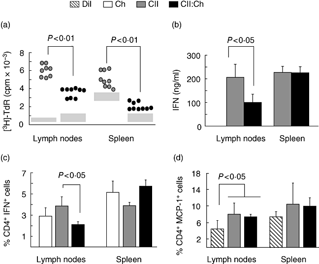
Fig. 4.

Evaluation of cellular response in draining lymph nodes and spleen of groups receiving either type II collagen (CII) or CII : chitosan. Mononuclear cells of draining lymph nodes and spleen (n = 7–9 rats per group) isolated 4 weeks after primary immunization were cultured for 72 h in the presence of 40 µg/ml CII. (a) Proliferative responses were assessed on the degree of [3H]-thymidine incorporation. Results are presented as counts per minute (cpm) of antigen-stimulated cells. Grey boxes represent the range of basal [3H]-thymidine incorporation; cpm values × 103 for chitosan group in lymph node 0·39 ± 0·102 (basal) and 5·24 ± 0·46 (CII-stimulated); in spleen 1·23 ± 0·24 (basal) and 4·41 ± 0·41 (CII-stimulated); (b) production of interferon (IFN)-γ by enzyme-linked immunosorbent assay in culture supernatants of mononuclear cells stimulated as described in (a); (c) percentage of CD4+ IFN-γ+ cells assessed by flow cytometry after stimulation in chitosan, CII- and CII : Ch-fed groups; (d) percentage of CD4+ monocyte chemotactic protein-1+ cells assessed by flow cytometry upon antigen stimulation in diluent, CII- and CII : Ch-fed groups.
