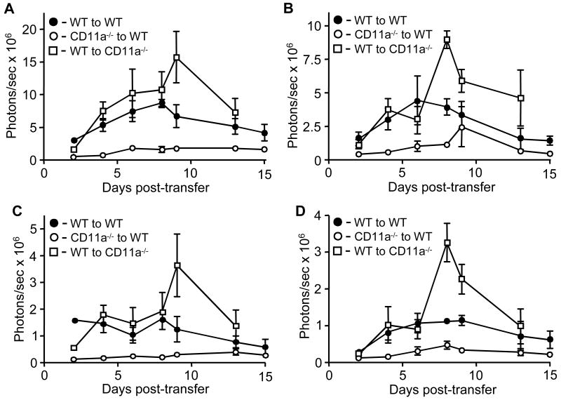
Figure 4.
Quantification of bioluminescence from cervical lymph nodes and brain and spinal cords during transferred EAE. Data were collected at days 2, 4, 6, 8, 9, 12, and 15 post-transfer from whole body ventral images (A), whole body dorsal images (B), cervical nodes (C) and the combination of brain and spinal cord (D) from all transferred EAE groups shown in Fig. 2. Results shown are the average of three to four mice per time point plotted as photons/sec versus days post transfer.

