


Figure 4. Acetylation-dependent extra-cellular localization of hsp90α.
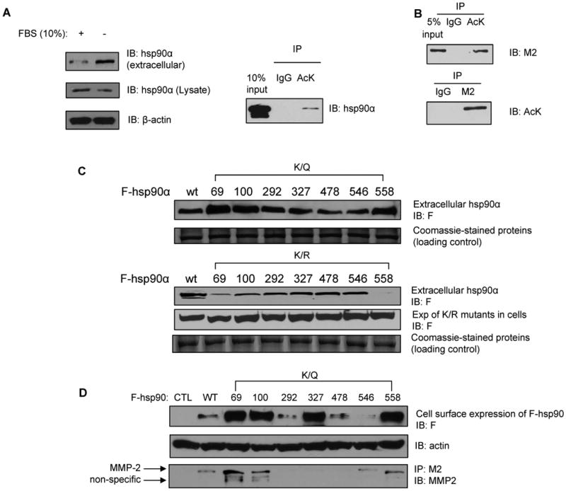
(A) Serum-starvation of T47D breast cancer cells promotes extra-cellular localization of hsp90α. Concentrated extra-cellular medium or cell lysates from T47D cells, which were either serum-starved or cultured in 10 % FBS, were immunoblotted with anti-hsp90α antibody. β-actin served as a loading control. (A, right panel and B) Under starvation, both endogenous (4A, right panel) and exogenous hsp90α (B) are secreted from T47D cells in the acetylated form. Extra-cellular hsp90α was immunoprecipitated with anti-AcK antibody and immunoblotted with either anti-hsp90α or M2 antibody. (C) In serum-starved T47D cells, K/R substitutions at K69, K100 and K558 decrease, while K/Q substitutions increase the level of extracellular hsp90α. Supernatants of serum starved T47D cells transfected with the indicated F-tagged hsp90α mutant constructs were concentrated and immunoblotted with anti-F antibody. Coomassie-stained non-specific proteins served as the loading control. (D) K/Q substitution affects hsp90 expression on cell surface. MB-231 cells transfected with the indicated constructs were cultured under serum-free condition for 24 hours and followed by the labeling of surface protein with biotinylation. Biotinylated hsp90 on cell surface was detected with anti-F antibody. Biotinylated actin on cell surface served as loading control. Supernatants of serum-starved MB-231 cells transfected with the indicated F-tagged K/Q hsp90α mutant constructs were also concentrated and immunoprecipitates with anti-M2 conjugated beads were immunoblotted with anti-MMP-2 antibody.