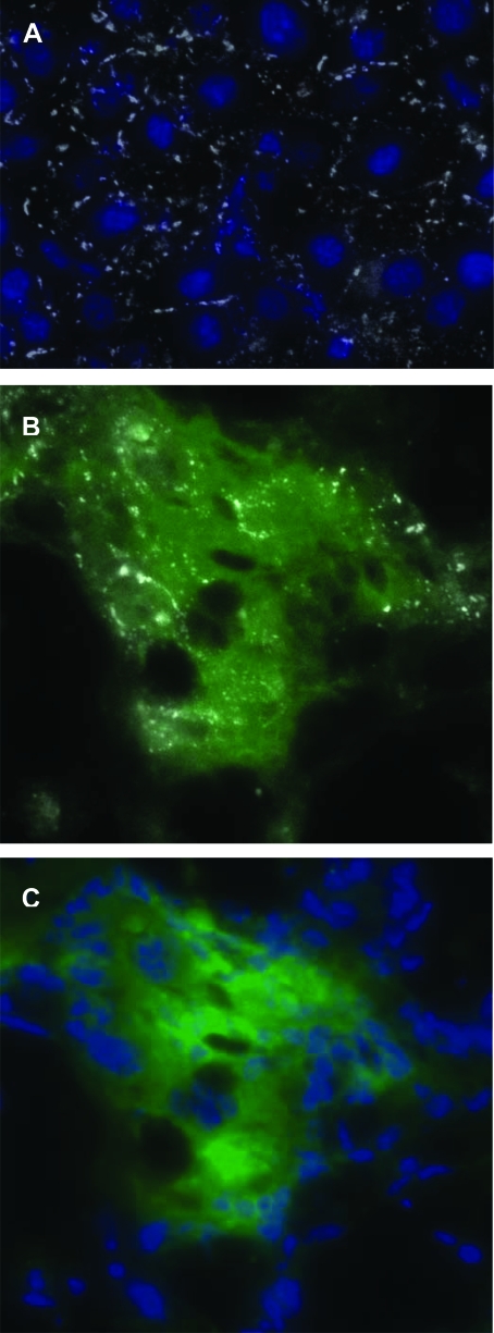
Figure 10.
Connexin-32 expression in a normal mouse liver and the liver of a recipient SCID mouse, treated as in Figure 2. A: Immunofluorescence image of a section of normal mouse liver stained with an anti-connexin-32 antibody (white) and with a nuclear dye (blue). B: A GFP+ colony in recipient SCID mouse liver (green), overlaid with the same section stained with an anti-connexin-32 antibody (white). C: Same section as in (B), showing the green fluorescence area with nuclear staining. Magnification = original ×400. Similar results were seen in the other treatment groups shown in Table 1 which had engraftment rates >50%.

