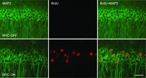
Figure 3.
DNA replication in the hippocampal neurons in MYC-On mice. Incorporation of BrdU during DNA replication is visualized using an anti-BrdU antibody (red). The number of BrdU-positive nuclei is dramatically increased in the hippocampal pyramidal neuron layer (CA1) of MYC-On mice. Double staining with anti-MAP2 antibody (green) clearly demonstrates that the BrdU-positive cells are neurons. In MYC-Off mice, virtually no BrdU-positive nuclei are observed. Scale bar = 50 μm.

