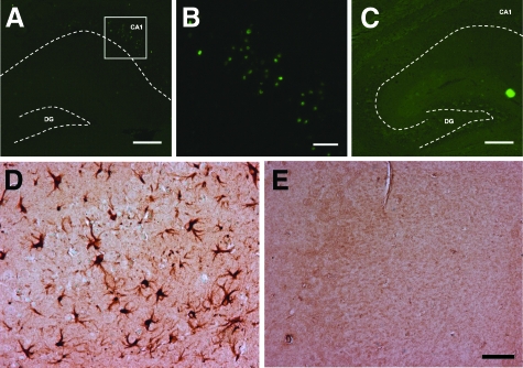
Figure 5.
Neurodegeneration and astrocytosis in the hippocampus in MYC-On mice. TUNEL-positive nuclei are specifically localized in the hippocampal CA1 region in MYC-On mice (A, B) but not in MYC-Off mice (C). B: High magnification of the framed portion in A. Immunocytochemistry with an anti-GFAP antibody demonstrates the extensive astrocytosis in the hippocampus in MYC-On mice (D) compared with MYC-Off mice (E). DG, dentate gyrus. Dotted lines outline the hippocampal subfields. Scale bars: 200 μm (A, C); 50 μm (B, D, E).

