Figure 10.
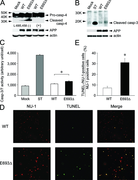
Increased apoptosis by the mutant Aβ. COS-7 cells were transfected with APPWT and APPE693Δ constructs and cultured for 2 days. A: Cell lysates were subjected to Western blotting with anti-caspase-4 antibody, in which the appearance of cleaved fragments of caspase-4 represents activation of caspase-4. Higher degrees of caspase-4 activation were observed in APPE693Δ-transfected cells, signals of which were completely abolished by the treatment of cells with 1 μmol/L γ-secretase inhibitor L-685,458. B: Cell lysates were subjected to Western blotting with an antibody to cleaved caspase-3, in which the appearance of the specific bands represents activation of caspase-3. As a positive control for apoptosis, mock-transfected cells were treated with 1 μmol/L staurosporine (ST) for 4 hours at 37°C. Higher degrees of caspase-3 activation were observed in APPE693Δ-transfected cells. C: Caspase-3 activity was measured in cells using the Caspase-Glo 3/7 assay kit, which includes luminogenic substrate for caspase-3/7. Again, higher luminescence was detected in APPE693Δ-transfected cells, indicating increased apoptosis of these cells. The columns and bars represent the means ± SD for four transfectants. *P = 0.0002 by unpaired Student’s t-test. D: Cells were fixed, permeabilized, and blocked as described in Figure 4, and then incubated with TUNEL label mix containing TUNEL enzyme (green). After washing, the cells were stained with NU-1 (red). E: The ratio of TUNEL-/NU-1-positive cells to NU-1-positive cells was calculated. The columns and bars represent the means ± SD for three experiments. *P = 0.0005 versus wild-type (WT) by unpaired Student’s t-test. In parallel with the increased accumulation of Aβ oligomers, stronger TUNEL-positive staining was observed in APPE693Δ-transfected cells, indicating increased DNA fragmentation, another sign of apoptosis, of these cells. Taken together, it was shown that the mutant Aβ causes ER stress-induced apoptosis.

