Figure 2.
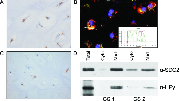
A: Central chondrosarcoma showing cytoplasmic dot-like accumulation of CD44v3 at immunohistochemistry, suggestive for Golgi retention. B: This was confirmed by IF using a Golgi-specific marker, 58K protein, in green, and CD44v3 in red, resulting in a yellow color when co-localization occurs. The white line indicates the position where the staining profile (inset) has been taken. C: Central chondrosarcoma showing nuclear staining of SDC2, which was found in 50% of chondrosarcomas in addition to cytoplasmic staining. D: Nuclear localization of SDC2 was verified by immunoblotting in two chondrosarcomas. In the isolated nuclear fraction heterochromatin protein 1γ was present, verifying the procedure for separating the nuclear from the cytoplasmic staining. Original magnifications, ×40.

