Figure 2.
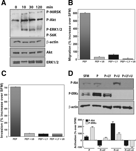
Proepithelin-mediated activation of the Akt and MAPK pathway promotes migration and invasion of DU145 cells. A: DU145 cells were serum-starved for 24 hours and stimulated with proepithelin (240 nmol/L) for 10, 30, and 120 minutes. The activation of p90RSK, Akt, ERK1/2, and S6 ribosomal protein (top to bottom) was analyzed by Western immunoblot using phosphoro-specific antibodies as described in Materials and Methods. The experiment shown is representative of three independent experiments. Migration (B) and invasion (C) experiments were performed on DU145 cells as described in Materials and Methods. U0126 (Calbiochem) was used at 10 μmol/L concentration and LY294002 was used at 30 μmol/L concentration. Values are expressed as percent increase over SFM condition (70 ± SD). D: Top: DU145 cells were serum-starved for 24 hours and then stimulated for 10 minutes with purified proepithelin (240 nmol/L). The activation of Akt and ERK1/2 in the presence of 10 μmol/L U0126 or 30 μmol/L LY294002 or the combination of the two was detected by immunoblot with anti-phospho-specific p90RSK and ERK1/2 (Cell Signaling). Samples were normalized using anti-β-actin polyclonal antibodies (Sigma-Aldrich). Bottom: Densitometric analysis of the Akt and ERK activation levels was performed using the Image J program (National Institutes of Health). Values in arbitrary units from three independent experiments are shown.

