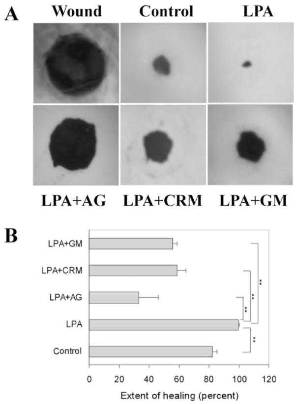
Figure 1.
Corneal epithelial wound healing in cultured porcine corneas. (A) Representative epithelial wound closure induced by LPA in cultured porcine corneas. A 4-mm diameter epithelial wound was made (Wound) and allowed to heal for 48 hours in MEM (Control), MEM containing LPA (5 μM), LPA plus AG1478 (1 μM), LPA plus CRM197 (10 μg/mL) or LPA plus GM6001 (50 μM). Wounded corneas were stained with Richardson staining solution to show the initial (Wound) and the remaining wound. (B) Changes in the extent of healing in cultured porcine corneas treated with different reagents. Coverage of the wound (0 for no migration and 100% for complete covering of the wound bed) was calculated as described in Materials and Methods. LPA accelerated corneal epithelial healing compared with control. However, the abilities of LPA to speed up healing were impaired in the presence of AG1478, CRM197, and GM6001 compared with the control (**P <0.01, one way ANOVA). Data are mean ± SD of at least six corneas from two or more independent experiments.

