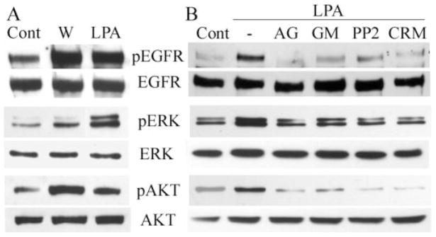
Figure 3.
Wound- and LPA-stimulated phosphorylation of EGFR, ERK, and AKT. (A) Growth factor–starved THCE cells (Cont) were extensively injured or were stimulated with 5 μM LPA for 15 minutes or (B) the cells were pretreated with AG1478 (1 μM), GM6001 (50 μM), PP2 (12.5 μM), or CRM197 (10 μg/mL) for 1 hour and then were stimulated with LPA (5 μM) for 15 minutes. Controls include cells without any stimulation (Cont) or cells stimulated with LPA alone (−). (A, B) Cells were lysed at the end of stimulation, and equal amounts of cell lysates were immunoprecipitated with EGFR antibody and immunoblotted by mouse pY99 antibody against tyrosine-phosphorylated proteins (pEGFR). The same membrane was stripped from the immunoreactivities and reprobed with EGFR antibody (EGFR) to assess the total amount of EGFR precipitated. The same cell lysates were subjected to Western blotting with anti–phospho-ERK1/2 (pERK) and -AKT using ERK2 and AKT levels, respectively, for proper protein loading.

