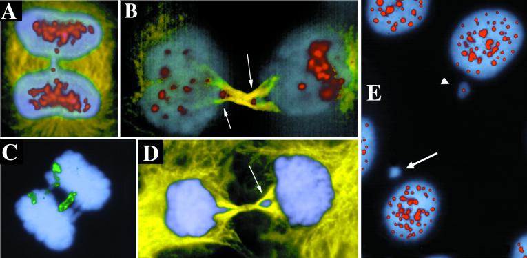
Figure 4.
Anaphase bridges containing centromeres and chromosome 11. Immunolabeling with Abs to tubulin (yellow), centromeres (red), and with DAPI (blue) and FISH with a chromosome 11 paint probe (green). (B) Arrows point to centromeres trapped in the forming midbody as these late telophase cells divide. (D) Arrow points to the trapped lagging chromosome excluded from the reforming nucleus of the cell on the right. (E) Some micronuclei are immunonegative for anti-centromere Abs. Arrow points to negative micronucleus, and arrowhead points to positive. Examples are from UPCI:SCC131.

