
Figure I.
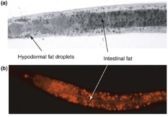
Fat staining of wild-type C. elegans. (a) Sudan Black staining of a fixed L4-stage larva. Black-stained fat droplets are visible in the intestinal and hypodermal cells. (b) Nile Red staining of a live L4-stage larva. Intestinal gut granules fluoresce red. In both pictures, anterior is to the left.