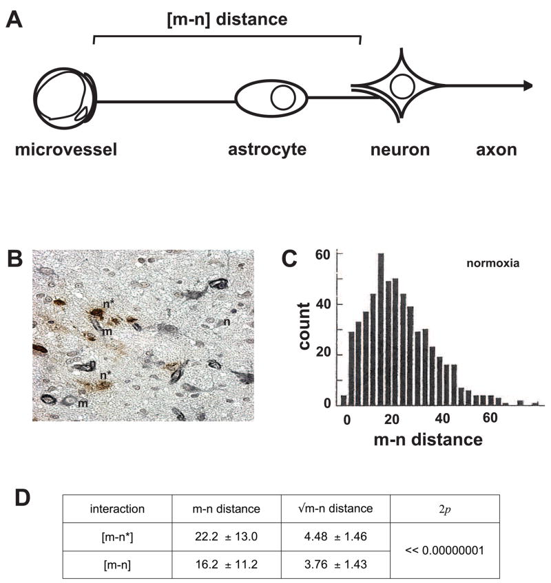
Figure 1.
Microvessel-neuron (m–n) inter-relationships based upon [m–n] distance distributions. A, the neurovascular unit and definition of [m–n] distance. B, indication of positions of microvessels (m) and neurons with evidence of dUTP incorporation (n*) and neurons without evidence of DNA scission (n) at 2 hours following middle cerebral occlusion (MCA:O) in the striatum of the non-human primate. C, untransformed data of [m–n] distances in the non-ischemic striatum of the non-human primate (in μm). D, neurons with evidence of injury (n*) at 2 hours MCA:O are at significantly greater distance from their nearest neighboring microvessel ([m–n*]) than those without evidence of injury ([m–n]). Data and figures from Mabuchi et al.(Mabuchi et al., 2005)

