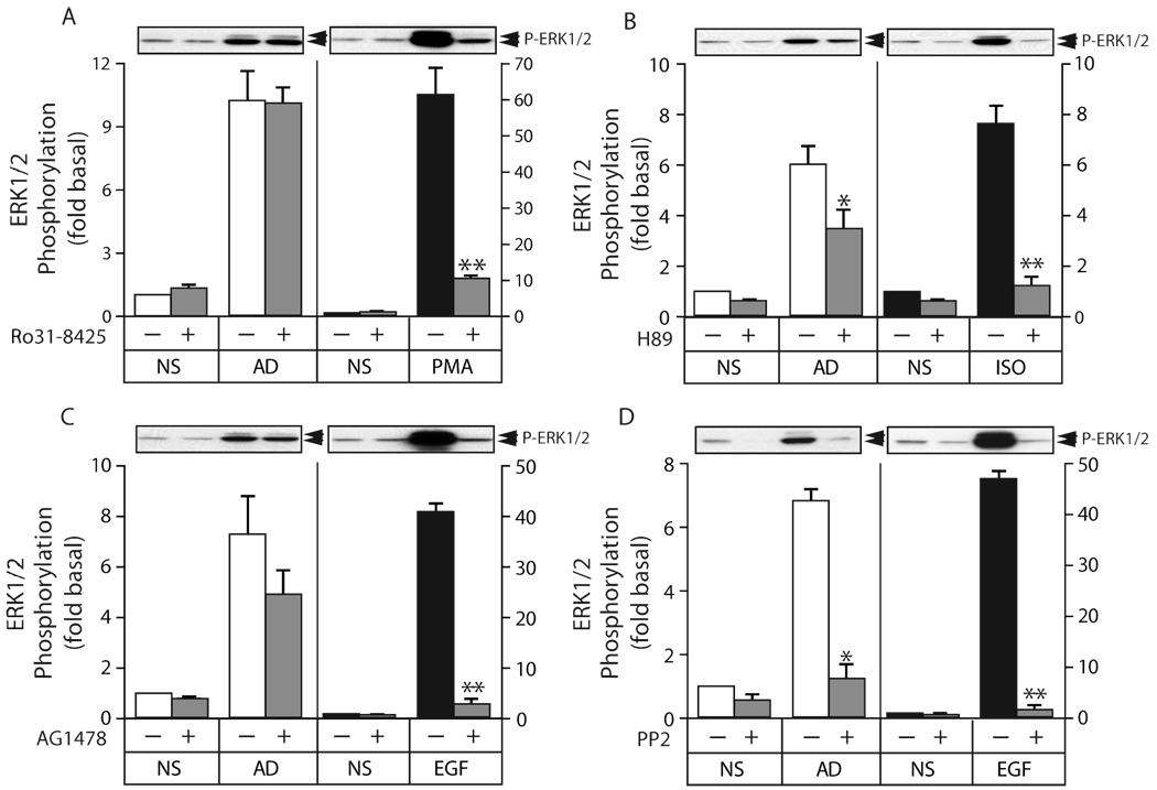
Figure 6. Effect of PKC, PKA, EGF receptor and Src inhibitors on adiponectin-mediated ERK1/2 activation.
A) Serum-deprived HEK293 cells were pretreated with the PKC inhibitor R031-8425 (1 µM) for 30 min prior to stimulation with full length adiponectin (8 µg/ml) or PMA (100 nM) for 5 min, after which phosphorylation of ERK1/2 was determined by immunoblotting. B) Cells were pretreated with the PKA inhibitor H89 (10 µM) for 30 min prior to stimulation with full length adiponectin (8 µg/ml) or isoproterenol (ISO; 1 µM) for 5 min, after which phosphorylation of ERK1/2 was determined. C) Cells were pretreated with the EGF receptor inhibitor AG1478 (200 nM) for 30 min prior to stimulation with full length adiponectin (8 µg/ml) or EGF (10 ng/ml) for 5 min, after which phosphorylation of ERK1/2 was determined. D) Cells were pretreated with the Src family kinase inhibitor PP2 (10 µM) for 30 min prior to stimulation with full length adiponectin (8 µg/ml) or EGF (10 ng/ml) for 5 min, after which phosphorylation of ERK1/2 was determined. In panels A–D, a representative phospho-ERK1/2 immunoblot is shown above a bar graph representing the Mean ± SEM from four independent experiments. ERK1/2 phosphorylation is expressed as the fold increase above the basal level in unstimulated cells not exposed to the inhibitor. * less than untreated, p<0.05; ** less than untreated, p<0.005.

