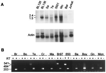
Figure 6.
Expression analysis of FcγRII in lymphoma. (A) Northern analysis of FcγRII expression in two Burkitt lymphoma cell lines (BL126, BL136) and four primary lymphoma samples (Sa, Te, 5501, Bar?), all with trisomy 1q compared with lymphoma case Mo (no 1q anomaly) and the T-cell line Jurkat. RNA was limited for three of these cases, so ≈10 μg instead of 20 μg was loaded for analysis (Sa, Te and Bar). The FcγRIIB probe (EC2/TM) does not distinguish between FcγRIIA, -B, or -C transcripts. (B) High level expression of the FcγRIIb2 isoform in a second case of follicular lymphoma (650) with t(1;22)(q22;q11). RT-PCR analysis of FcγRIIB expression in lymphoma case 650 compared with nine other lymphomas of diverse histological subtypes with trisomy 1q (Br, Bo, Te, Cr, and Ma) or without trisomy 1q (Br97, Ba, Boy, Gn and Mon). The RT control is shown for the FcγRIIB-specific RT-PCR only.

