
Figure 3. Schematic of bicelle structure and 31P chemical shift spectra of DMPC:DMPG:DHPC bicelles containing IAPP.
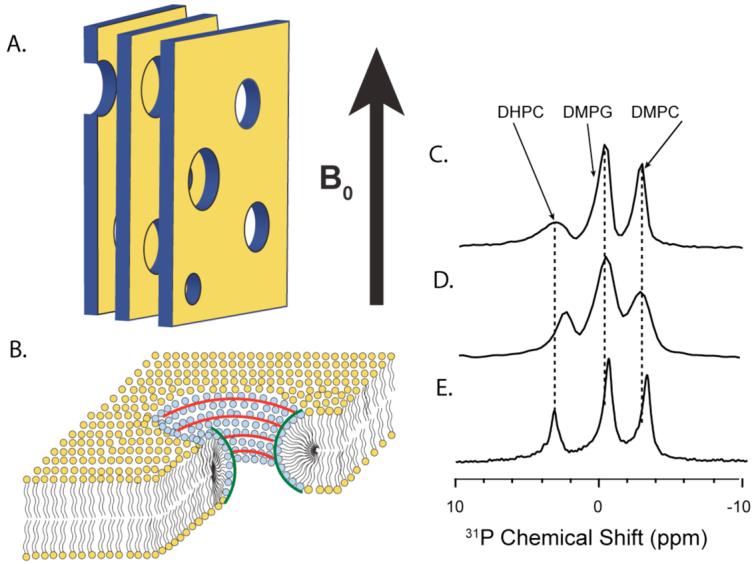
(A) A cartoon depiction of magnetically-aligned bicelles in the lamellar phase showing the parallel bicelle lamellae composed of DMPC and DMPG and the perforations composed of DHPC. The large, static magnetic field of the NMR spectrometer is indicated. (B) Zoomed in cartoon depiction of the bicelles, showing the regions of positive and negative curvature. (C) The 31P NMR spectrum of the pure bicelle sample. (D) The 31P NMR spectrum of the rIAPP1-19 bicelle sample. (E) The 31P NMR spectrum of the rIAPP1-37 bicelle sample.