
Figure 4.
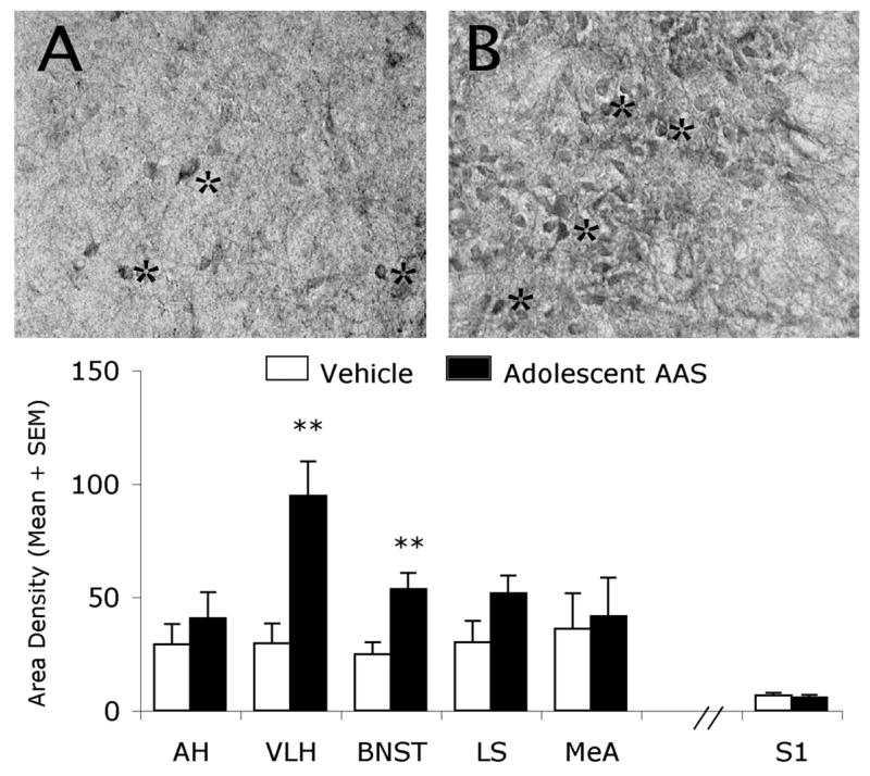
Brightfield photomicrographs of a coronal section through the Syrian hamster hypothalamus. Shown are GluR1-containing cells (astericks) within the ventrolateral hypothalamus of Vehicle- (A) and AAS- (B) treated hamsters. Graph represents density of GluR1-immunostaining (neuropil and somata) in select brain regions of Vehicle (White Bars) and AAS (Black Bars) treated hamsters. ** p< 0.01; Student’s t-test, two-tailed.