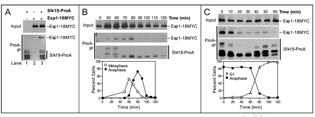
Figure 1.
The Esp1-Slk19 interaction is cell cycle regulated. (A) Western blots showing that Esp1-18MYC coimmunoprecipitates with Slk19-ProA. The top panel (Input) shows the amount of Esp1-18MYC in whole cell extracts. The bottom panel (ProA-IP) shows the amount of Esp1-18MYC coimmunoprecipitated with Slk19-ProA (lane 3). ProA-tagged proteins were detected in anti-MYC immunoblots because ProA binds to primary and secondary antibodies. The following strains were used (from left to right): A8196, A3072, A8444. (B) Cells carrying Slk19-ProA and Esp1-18MYC fusions (A8444) were arrested in G1 in YEPD with alpha factor (5 µg/ml) at room temperature. After 2.5 hours, cells were washed with 10 volumes YEP and released into pheromone-free YEPD media at room temperature. After 100 minutes, alpha factor (10 µg/ml) was re-added to prevent cells from entering a new cell cycle. Samples were taken at the indicated times to determine the ability of Esp1 and Slk19 to interact (top) and the percentage of metaphase (open circles) and anaphase (closed squares) spindles (bottom). The Western blots show the amount of Esp1-18MYC in whole cell extracts (Input, top), the amount of Esp1-18MYC coimmunoprecipitated with Slk19-ProA (ProA-IP, middle), and the amount of Slk19-ProA immunoprecipitated (ProA-IP, bottom) at the indicated times after release from the G1 arrest. (C) cdc15-2 cells carrying Slk19-ProA and Esp1-18MYC (A11605) fusions were arrested in G1 in YEPD with alpha factor (5 µg/ml) for 2.5 hours at room temperature. Cells were washed with 10 volumes YEP and released into pheromone-free YEPD media at 37°C for 2.5 hours to arrest cells in anaphase. Cells were then released into YEPD media at room temperature in the presence of alpha factor (5 µg/ml) for cells to exit from mitosis and arrest in the following G1 stage. Samples were taken at the indicated times to determine the ability of Esp1 and Slk19 to interact (top) and the percentage of anaphase closed squares) and G1 (open triangles) spindles (bottom). The Western blots show the amount of Esp1-18MYC in whole cell extracts (Input, top), the amount of Esp1-18MYC coimmunoprecipitated with Slk19-ProA (ProA-IP, middle), and the amount of Slk19-ProA immunoprecipitated (ProA-IP, bottom) at the indicated times after release from the late anaphase arrest.

