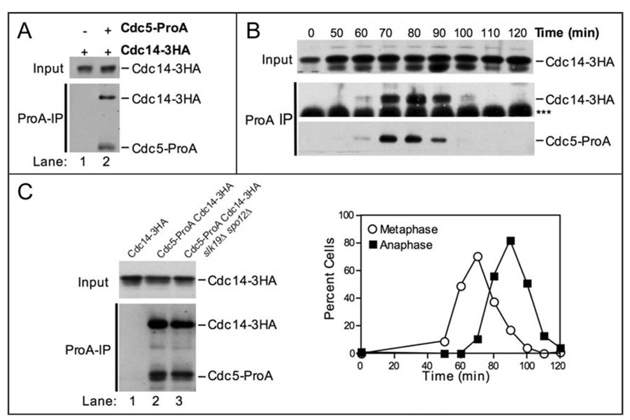
Figure 3.
Cdc5 physically interacts with Cdc14. (A) Western blots showing that Cdc14-3HA coimmunoprecipitates with Cdc5-ProA from whole cell extracts. The top panel (Input) shows the amount of Cdc14-3HA in whole cell extracts. The bottom panel (ProA-IP) shows the amount of Cdc14-3HA coimmunoprecipitated with Cdc5-ProA (lane 2). The following strains were used (from left to right): A1411, A7216. (B) Immunoprecipitates of Cdc5-ProA from the experiment shown in Figure 2C were probed with an HA.11 antibody to determine the amount of Cdc14-3HA coimmunoprecipitated. The Western blots show the amount of Cdc14-3HA in whole cell extracts (Input, top), the amount of Cdc14-3HA coimmunoprecipitated with Cdc5-ProA (ProA-IP, middle), and the amount of Cdc5-ProA immunoprecipitated (ProA-IP, bottom) at the indicated times after release from the G1 arrest. The bottom panel is a lighter exposure of the panel shown in Figure 2C. *** denotes the IgG heavy chain. The graph showing the percentage of cells in metaphase and anaphase is the same as that shown in Figure 2C. (C) Western blots showing that Cdc14-3HA coimmunoprecipitates with Cdc5-ProA in the absence of SLK19 and SPO12. The top panel (Input) shows the amount of Cdc14-3HA in whole cell extracts. The bottom panel (ProA-IP) shows the amount of Cdc14-3HA coimmunoprecipitated with Cdc5-ProA. The following strains were used (from left to right): A1411, A7216, A12424.

